FIGURE 6.
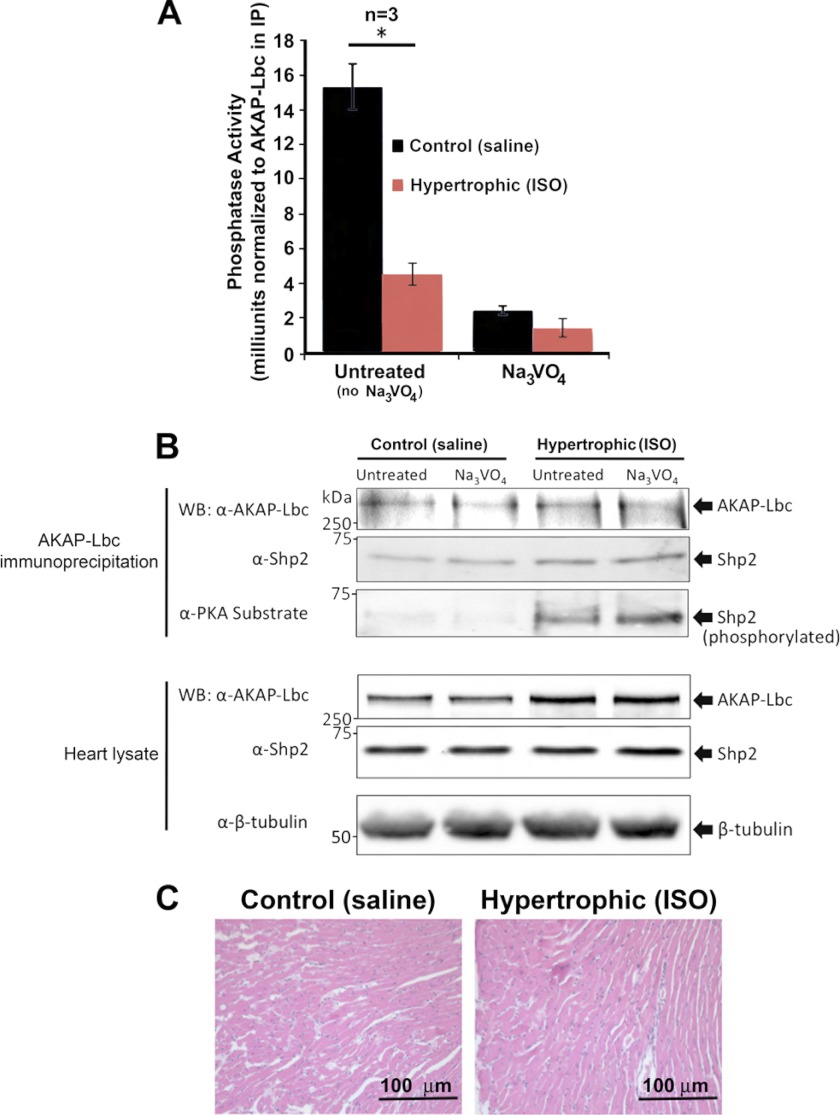
Shp2 activity is decreased in isoproterenol-induced hypertrophic hearts. A, AKAP-Lbc complexes were isolated from healthy (Control; saline-treated) and hypertrophic (ISO-treated) mouse heart extract by immunoprecipitation using anti-AKAP-Lbc antibody. Immunoprecipitates were washed, and PTP activity was measured as described previously. Results presented show PTP activity ± S.E. measured in triplicate from three hypertrophic hearts and three age-matched control healthy hearts. Control IgG IP background activity has been subtracted, and the PTP activity was normalized to AKAP-Lbc expression in the immunoprecipitation. Untreated refers to no Na3VO4. B, Western blotting of AKAP-Lbc and associated Shp2 levels in the IPs used for PTP assay. Bottom panels show levels of AKAP-Lbc and Shp2 in heart lysate used for IP. C, histological analysis of mouse hearts corresponding to samples used for PTP assay. Hematoxylin/Eosin staining of left ventricle sections indicates isoproterenol-induced hypertrophic myocytes (Table 1). Echocardiography characteristics indicate ventricular dilation and hypertrophy.

