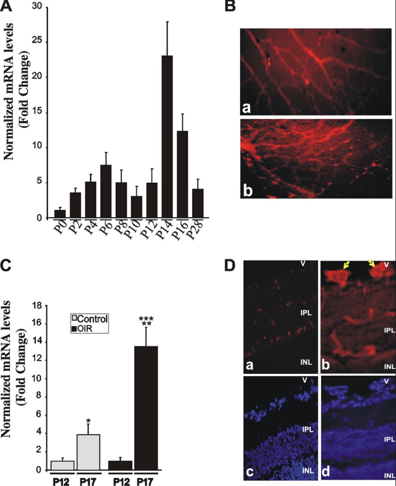
FIGURE 1.
Expression and tissue localization of CTGF/CCN2 during normal retinal vessel development and in response to OIR. A, room air mouse pups were raised under normal light and temperature conditions, sacrificed at the indicated time periods, and retinas dissected. CTGF/CCN2 mRNA levels were quantified by qPCR and normalized to those of 18 S rRNA. The ΔΔCt values are the average of determinations from tissue samples obtained from 4 to 9 animals each measured in triplicate. B, immunohistochemical localization of the CTGF/CCN2 protein in flat-mount preparations of retinas at P12. All retinal preparations were fixed in 4% paraformaldehyde and permeabilized prior to immunostaining with anti-CTGF/CCN2 antibody. Immunoreactivity to CTGF/CCN2 in the superficial and deeper retinal vessels is shown in panels a and b, respectively. Magnification, ×40. C, expression pattern of CTGF/CCN1 mRNA following the hyperoxic (P7 to P12) and ischemic (P12 to P17) phases of OIR as determined by qPCR. CTGF/CCN2 mRNA levels were normalized to those of 18 S rRNA. Data are means ± S.E. (n = 4 animals). *, p < 0.05 versus P12/Control. **, p < 0.001 versus P12/OIR. ***, p < 0.05 versus P17/Control. D, immunolocalization of CTGF/CCN2 (panels a and b) in 10-μm-thick cross-sections of retinas from P17 control (panel a) and OIR (panel b) retinas. Nuclear staining with DAPI of cross-sections (panels a and b) is shown in panels c and d, respectively. Arrows indicate CTGF/CCN2 immunostaining in neovascular tufts protruding luminally in the vitreous. V, vitreous; IPL, inner plexiform layer; INL, inner nuclear layer. Magnification, ×40.

