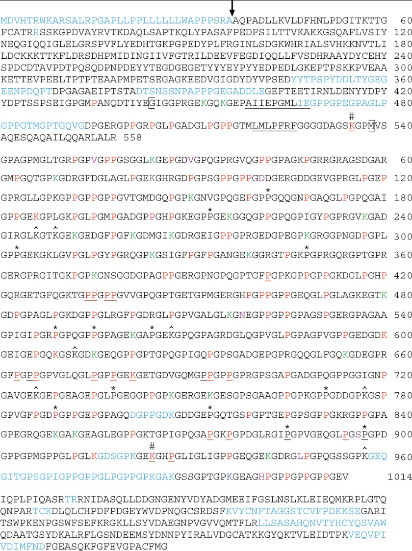
FIGURE 3.
Hydroxylated amino acid residues and glycosylated Hyl residues of recombinant human pro-α1(V) collagen chains produced in 293-EBNA human embryonic kidney cells. Sequence coverage was 90% of the entire pro-α1(V) chain and 96% of the COL1 domain. Within the COL1 domain 98 Y-position Hyp, 9 Gly-Hyp-Hyp X-position Hyp, 1 Gly-Hyp-Val, 1 Gly-Hyp-Ala, 1 Gly-Hyp-Thr, 7-Hyl, and 23 Glc-Gal-Hyl residues were identified. Red, hydroxylated, non-glycosylated residues; green, Glc-Gal-Hyl residues; dark blue, Gal-Hyl residue; purple, residue found as both Glc-Gal-Hyl and Gal-Hyl. Underlined, hydroxylated/glycosylated residues mapped in previous studies (4, 32). Sequences not identified in the course of MS analysis are in light blue. The seven COL1 amino acid residues that differ between human and bovine are orange. *, 12 Y-position Pro residues hydroxylated in human or bovine α1(V) COL1 domain but not the other. #, Hyl residues identified by Wu et al. (4) as being involved in interchain covalent cross-links. ^, Lys residues hydroxylated in bovine placenta α1(V) but not in human recombinant pro-α1(V). A vertical arrow marks the site of proteolytic removal of the signal peptide, as previously determined by Edman degradation NH2-terminal amino acid sequencing (40) and as confirmed by MS analysis in the present study. The brackets indicates limits of the small COL2 hypothetical triple helical domain, in which interruptions of Gly-X-Y repeats are underlined. Amino acid residues NH2-terminal to the COL1 domain are numbered 1 to 558, starting with the initial Met residue of the signal peptide. Amino acid residues of the COL1 domain are numbered 1 to 1014, for easy comparison to the similarly numbered residues of the bovine α1(V) COL1 domain in Fig. 1. Amino acids of the COOH-propeptide are shown for the sake of completeness, but are not numbered, due to lack of identified hydroxylated residues.

