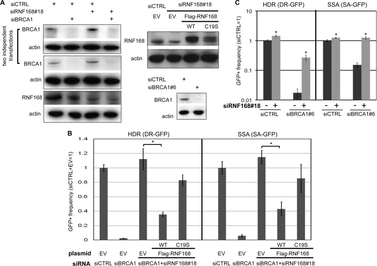
FIGURE 2.
Transient RNF168 expression inhibits HR in cells co-depleted of BRCA1 and endogenous RNF168. A, immunoblot analysis showing RNAi depletion of BRCA1 and RNF168, and expression of FLAG-RNF168. Shown are immunoblot signals for BRCA1, RNF168, and actin for cells treated with siRNAs used in B and C. Also shown (upper right) are immunoblot signals for RNF168 and actin for cells after co-transfection with siRNA along with the expression vectors that are used in B for FLAG-RNF168 WT, FLAG-RNF168 C19S (a RING domain mutant), or control EV. B, depletion of RNF168 by a distinct single siRNA (#18) suppresses HR defects in siBRCA1-treated cells, which is inhibited by transient expression of RNF168. U2OS reporter cell lines were examined as in Fig. 1C, except using siRNF168#18 and including siRNF168#18-resistant expression vectors for FLAG-RNF168 WT, FLAG-RNF168 C19S, or control EV during the I-SceI transfection. Shown are the frequencies of GFP+ cells for the HDR and SSA reporter cell lines and each siRNA treatment, relative to siCTRL and EV. *, p < 0.0001 (n = 3). C, repair analysis using a distinct siBRCA1. U2OS reporter cell lines were examined as in Fig. 1C, using siBRCA1#6 and siRNF168#18. Shown are the frequencies of GFP+ cells for the HDR and SSA reporter cell lines for each siRNA treatment, relative to siCTRL. *, p ≤ 0.0001 (n = 6).

