FIGURE 4.
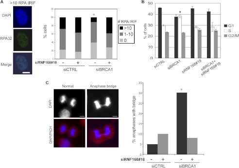
Depletion of RNF168 suppresses RPA IRIF and anaphase chromosome separation defects caused by BRCA1 silencing. Prior to examining RPA IRIF, cell cycle, and anaphases, U2OS cells were treated with siCTRL or siBRCA1, along with (+) or without (−) siRNF168#18, as in Fig. 2. A, depletion of RNF168 suppresses the decrease in RPA IRIF caused by siBRCA1 treatment. Cells 2 days after transfection were treated with 10 Gy of IR (Cs137) and allowed to recover for 4 h prior to fixation and immunostaining with RPA (RPA32 subunit) antibodies. Shown is a representative siCTRL-treated cell with >10 RPA IRIF, and the percentage of cells with 0, 1–10, and >10 RPA IRIF for each siRNA treatment. *, p < 0.0001 for number of cells >10 RPA IRIF versus all other siRNA treatments using Fisher's exact test (n > 200 accumulated from two independent transfections). B, treatment with siBRCA1 causes a modest shift of G1 to S phase cells. Shown are the percentages of cells in G1, S, and G2/M for each siRNA condition, based on co-staining for BrdU and propidium iodide. *, p = 0.0008 versus siCTRL (n ≥ 3). C, depletion of RNF168 suppresses the anaphase bridges caused by siBRCA1 treatment. Shown are representative images of a normal anaphase, and an anaphase bridge co-stained with PICH and DAPI from siBRCA1-treated cells. Bars, 10 μm. Shown is the percentage of bridges in late anaphase for each siRNA treatment, scored using DAPI staining. This frequency was determined from analyzing over 100 anaphases for each siRNA treatment, which were accumulated from several independent transfections (n ≥ 3). *, p < 0.0006 versus all other siRNA treatments using Fisher's exact test.

