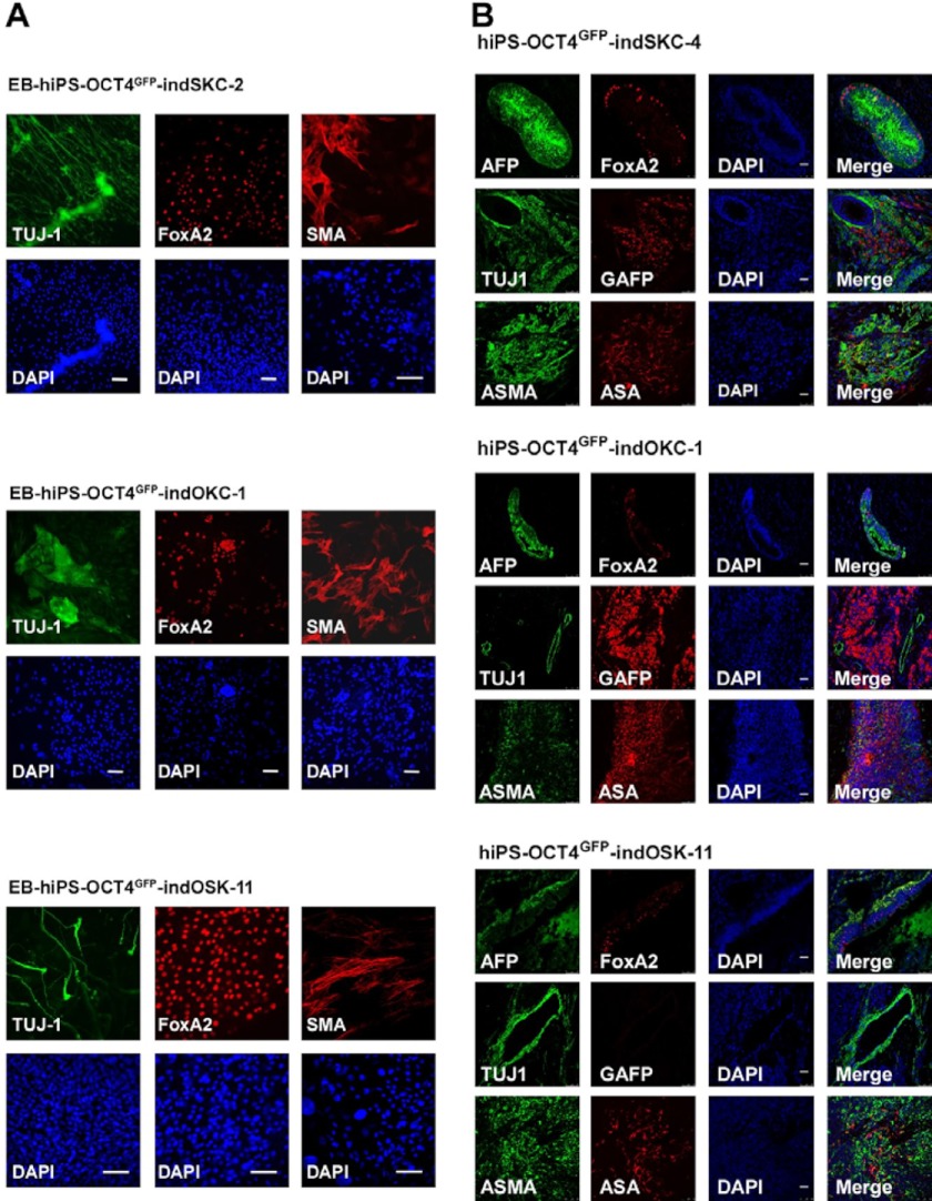
FIGURE 3.
hiPS-OCT4GFP–ind cells are pluripotent. A, embryoid bodies derived from the indicated hiPS-OCT4GFP–ind cells lines were differentiated for 15 days. Immunofluorescence was performed for specific differentiation markers from the three embryonic germ layers as defined by the expression of mesodermal (α-smooth muscle actin (SMA)), ectodermal (TUJ1), and endodermal (FoxA2) markers. Scale bar = 50 μm. B, teratoma formation was assessed by injection of the hiPS-OCT4GFP–ind cells lines into the testes or kidney of SCID mice. Immunofluorescence analysis demonstrate the existence of the three main embryonic germ layers as defined by the expression of specific endodermal (α-fetoprotein (AFP) and FoxA2), ectodermal (TUJ1 and glial fibrillary acidic protein (GFAP), and mesodermal (ASMA and α sarcomeric actin (ASA)) markers. All images were obtained from the same tumor. DAPI was used to visualize the nuclei. Scale bar = 50 μm.

