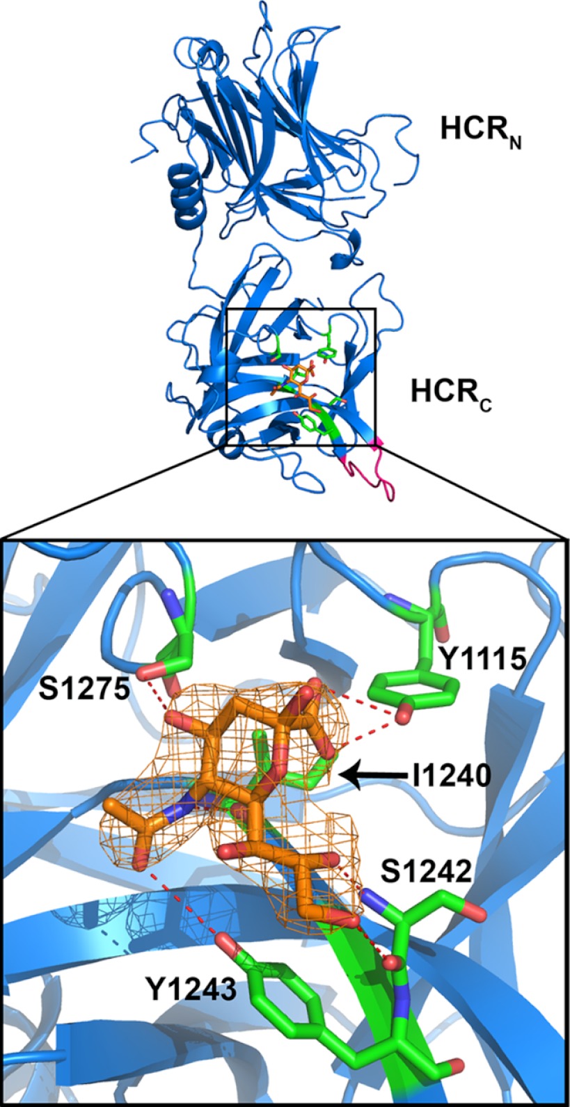
FIGURE 2.
A novel sialic acid binding pocket within HCR/D-C(GBL/C). The crystal structure of HCR/D-C(GBL/C) was solved in complex with sialic acid and is shown here (PDB code, 4FVV). Sialic acid is shown in orange. Residues of HCR/D-C(GBL/C) in contact with sialic acid are shown in green and define the GBP2. The N (HCRN)- and C (HCRC)-terminal subdomains of the HCR are labeled. GBL/C is shown in pink. The boxed area is expanded and shown (bottom). The sialic acid electron density is shown. [2Fo − Fc] contoured at 1σ. The residues TYR-1115, Ile-1240, Ser-1242, Tyr-1243, and Ser-1275 in HCR/D-C form direct H-bonds with sialic acid (red dashed lines). Nitrogen atoms are shown in blue and oxygen atoms in red.

