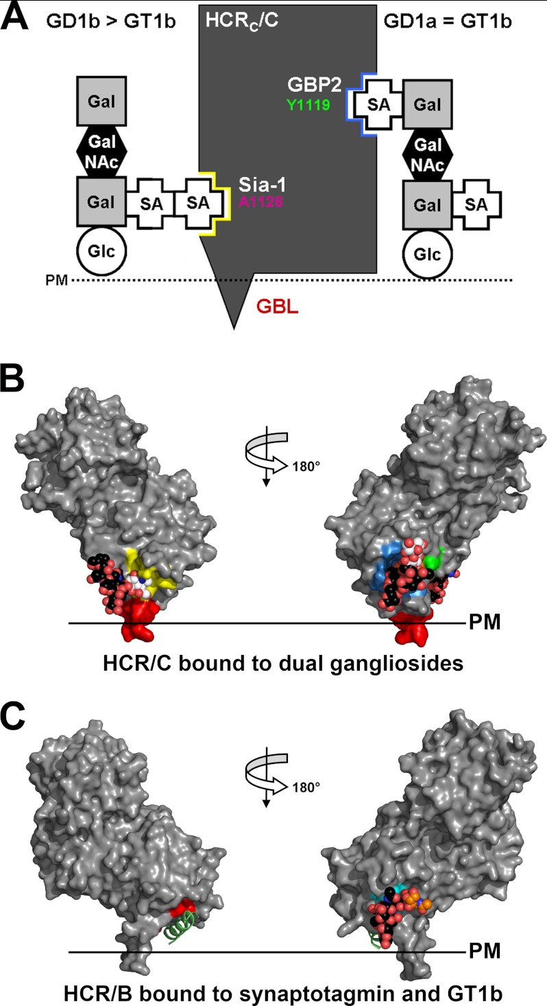
FIGURE 7.
BoNT/C binds dual gangliosides. A, schematic showing how BoNT/C binds ganglioside was generated based on the results of the mutagenesis study from Fig. 4. B, a manually generated model for HCR/C bound to dual ganglioside molecules (bound sialic acid moiety is shown in white, and the remainder of the ganglioside in black) was built by docking the sia7 from GD1b into the Sia-1 site using the Sia-1 molecule as a reference (PDB code, 3R4S) (26). The sia5 from GD1a was modeled into the GBP2 using the sialic acid bound by the HCR/D-C(GBL/C) as a reference. Ganglioside binding sites are colored as described in the legend for Fig. 3. Binding of dual gangliosides positioned HCR/C to interact with the plasma membrane as shown with the GBL (red), penetrating the plasma membrane (PM) lipid bilayer. C, HCR/B bound to dual receptors: synaptotagmin and GT1b. Alignment of HCR/B with the synaptotagmin peptide (green; PDB code, 2NM1) (15, 16) and GT1b bound to HCR/A (PDB code, 2VU9) (45) are shown. Residues that contact the synaptotagmin peptide and GT1b are shown in red and cyan, respectively.

