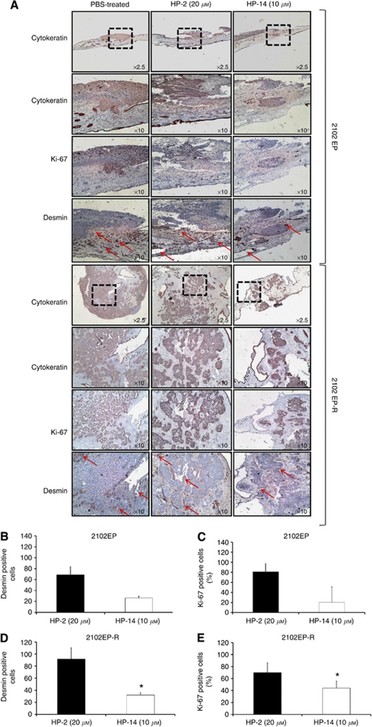
Figure 6.
Immunohistochemical analysis of CAM tumours. (A) Paraffin-embedded cisplatin-sensitive (upper panel) and cisplatin-resistant (lower panel) tumours were subjected to immunohistochemistry with specific antibodies directed against human cytokeratin, Ki-67 and desmin. (B, D) Quantification of HP-induced inhibition of blood vessel formation (desmin immunostaining) of cisplatin-sensitive (B) and -resistant (D) tumours. Data are given as the percentage of desmin-positive cells after treatment with HP-2 or HP-14, as compared with untreated controls. (C, E) Quantification of HP-induced changes in Ki-67 staining in cisplatin-sensitive (C) and -resistant tumours (E). Immunoreactivity was quantified by counting positive cells. For each slide, 16 fields were randomly chosen and, using a defined field area (1.72 mm2), all positive cells per field were counted. Results represent the mean±s.e.m. of three tumours. Data were analysed by Student’s t-test. *P<0.05 compared with controls.

