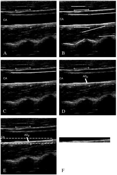
Figure 4.
Automated carotid identification (Stage-I) by CALEX 3.0. A) Original cropped image; B) line segments; C) line segments corresponding to the near and far adventitia layers obtained through validation and classification;25 D) final profile of the far adventitia (ADF); E) determination of a Guidance Zone in which segmentation is performed (white dashed rectangle); F) extracted Guidance Zone of the distal wall.

