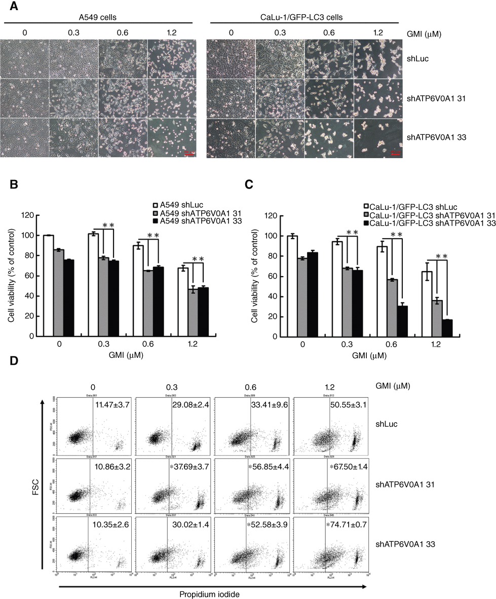
Figure 5.

Effect of ATP6V0A1 silencing on GMI-induced cell death in non-small cell lung cancer cells. (A) A549 shLuc, A549 shATP6V0A1, CaLu-1/GFP-LC3 shLuc and CaLu-1/GFP-LC3 shATP6V0A1 cells were treated with GMI for 48 h. Cell morphology was observed under an inverted microscope. Scale bar indicates 100 µm. (B) A549 shLuc, A549 shATP6V0A1, (C) CaLu-1/GFP-LC3 shLuc and CaLu-1/GFP-LC3 shATP6V0A1 cells (5 × 10³ cells per well of 96-well plate) were treated with GMI (0, 0.3, 0.6 and 1.2 µM) for 48 h. MTT assay was used to estimate the cell viability. The symbol (**) indicates P < 0.0001 by Student's t-test. (D) A549 shLuc, A549 shATP6V0A1 cells (2 × 10⁵ cells per well of 6-well plate) were treated with GMI (0, 0.3, 0.6 and 1.2 µM) for 48 h, followed by staining with propidium iodide. The fluorescence-activated cells were analysed by flow cytometry. The symbol (*) indicates P < 0.05 by Student's t-test.
