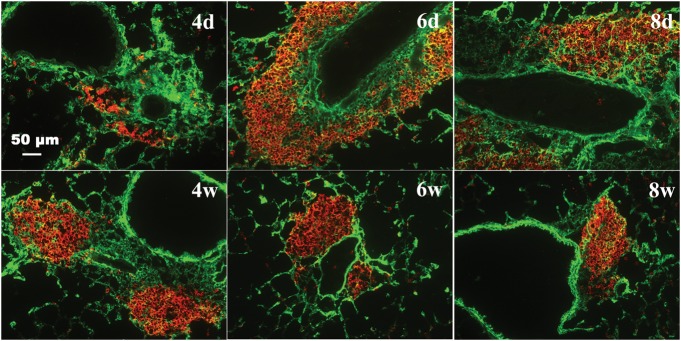
Fig. 12.
Co-localization of HA and B-lymphocytes in the lung during acute and chronic stage of antigen exposure. Lung sections were stained with HABP [green (full colour versions of figures are available online)] and B-lymphocytes (red), which demonstrate B cells embedded in the HA matrix (magnification bar 50 μm). Selected time points were used in this figure, and additional low magnification fields are shown in Supplementary data, Figure S8.

