
Figure 1. αvβ6 integrin expression in livers of normal and fibrotic rats and mice, and in various liver cells.
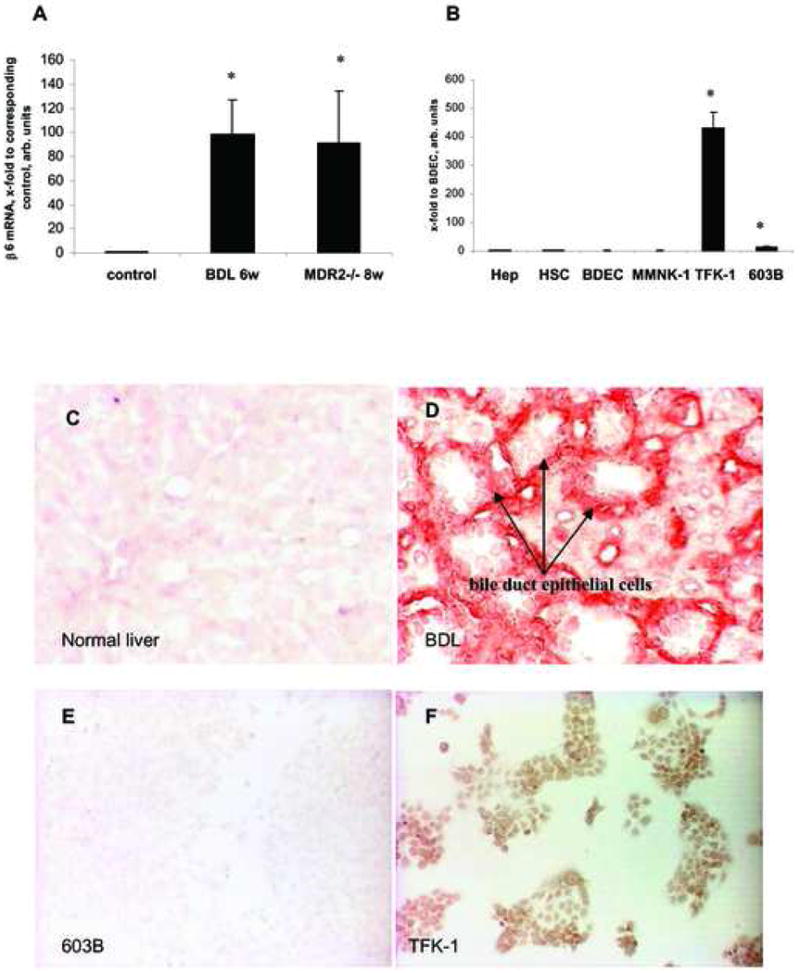
Rate limiting β6 transcript levels as quantified by real time PCR in: (A) Rat livers after 6 weeks of BDL and livers of 8 wk old MDR2-/- mice, both normalized to their age-matched sham-operated and wildtype controls, respectively; data in the MDR2-/-mice are from ref.29; (B) normal rat hepatocytes (Hep), rat hepatic stellate cells (HSC), human bile duct epithelial cells (BDEC), normal human (MMNK-1) or mouse (603B) cholangiocyte cell lines, and activated tumor-derived TFK-1 cholangiocytes, normalized to β2MG mRNA (x-fold increase relative to normal human BDEC). Means±SD; *p<0.05 vs. the corresponding control group.
αvβ6 immunostaining of liver sections from sham-operated rats (C), rats with BDL (D) or of normal mouse (603B) (E) and activated human (TFK-1) (F) cholangiocytes. αvβ6 protein is absent from normal livers and 603B cholangiocytes, but highly expressed on proliferating bile duct epithelial cells (red) and TFK-1 cholangiocytes (brown). Shown are representative images (magnification 40×).