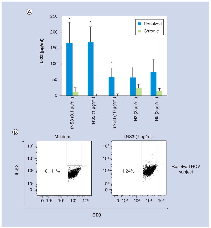
Figure 3. T-cell production of IL-22 was significantly different between resolved and chronic HCV subjects.
(A) Peripheral blood mononuclear cells from resolved (n = 8) and chronic (n = 8) subjects were incubated with either medium, rNS3 (0.1, 1 and 10 μg/ml), H3 (3 μg/ml), H5 (3 μg/ml) or phytohemagglutinin (2 μg/ml). Cell culture supernatants were collected 48 h after the addition of antigens, mitogen or medium. Data are normalized to medium. (B) Representative flow cytometry dot plots of CD3+ IL-22+ expression in a resolved HCV subject’s cells at 48 h post-antigen stimulation. Flow cytometry data are representative of four different experiments.
*p < 0.05, Student’s paired t test.
rNS3: Recombinant NS3.

