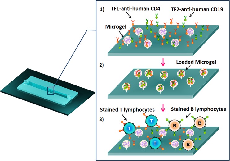
Figure 3.
Schematic representation of double cell staining within microgels functionalized microchannel. (1) Uptake of TF-labeled MAbs into the covalently immobilized PMAA microgels within the microchannel specific for the detection of B and T cells. (2) PMAA microgels microchannel loaded with TF1-antiCD4 and TF2-antiCD19 antibodies. (3) A defined cell mixture of two human lymphoblastoid cells (TK6 and SUP-T1 cells) was incubated within the microchannel. The physiological pH of cells suspension induces the pH-triggered cell staining.

