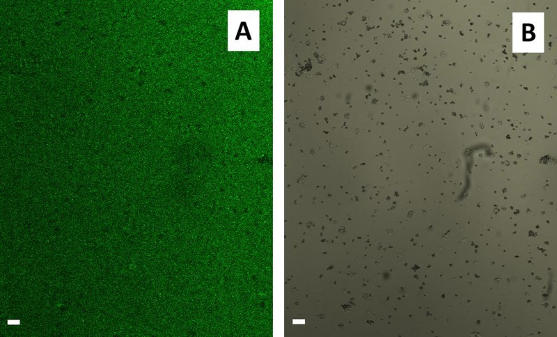
Figure 5.
Confocal microscopy images of the unloaded PMAA microgels microchannel after the release process of TF1-antiCD4 and TF2-antiCD19 antibodies induced by the passage of MES buffer (1.0 mM) at physiological pH (pH 7.8) within it. (a) Fluorescence image of the empty microgels functionalized microchannel. (b) The corresponding image acquired under bright field exposure. The scale bar was 20 μm.

