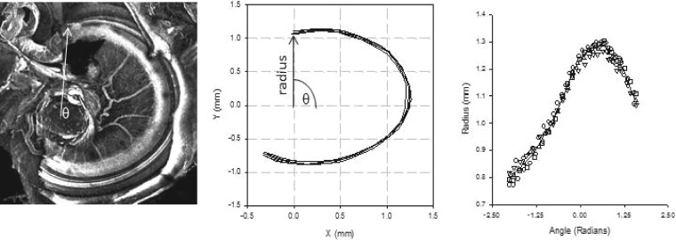
Figure 8.
A 4× image of the basal turn of the gerbil cochlea obtained with a confocal microscope. The arrow points to the beginning of the cochlear partition but also serves as the radius of the cochlear partition rotated around angle theta (θ), located in the center of the modioulus (left panel). Middle panel shows the result of five tracings of the cochlear partition. The coordinates of these tracings were converted to polar coordinates and fit with a cubic spline (right panel).

