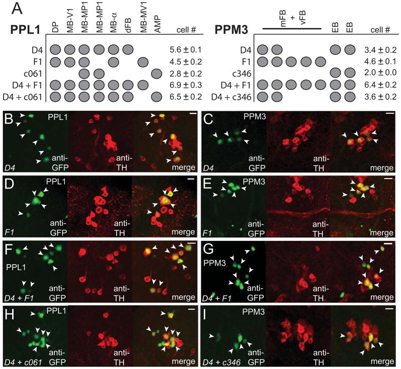
Figure 4. A Single Neuron from the PPL1 Subgroup, which Projects to the Dorsal Fan-shaped Body, Promotes Wakefulness.
(A) Identities of the TH+ cells in the PPL1 and PPM3 clusters in the indicated Gal4 driver lines. Mean cell counts for PPL1 and PPM3 are shown for TH-D4-Gal4 (n=13 hemispheres), TH-F1-Gal4 (n=14), c061-Gal4 (n=5), c346-Gal4 (n=3), TH-D4-Gal4/TH-F1-Gal4 (n=11), c061-Gal4; TH-D4-Gal4 (n=8), and c346-Gal4; TH-D4-Gal4 (n=9) flies expressing GFP-nls. (B–I) Single confocal sections showing the TH+ cells in the PPL1 or PPM3 clusters for the indicated driver lines expressing GFP-nls. Arrowheads indicate GFP+ TH+ cells. Scale bar denotes 10 μm.

