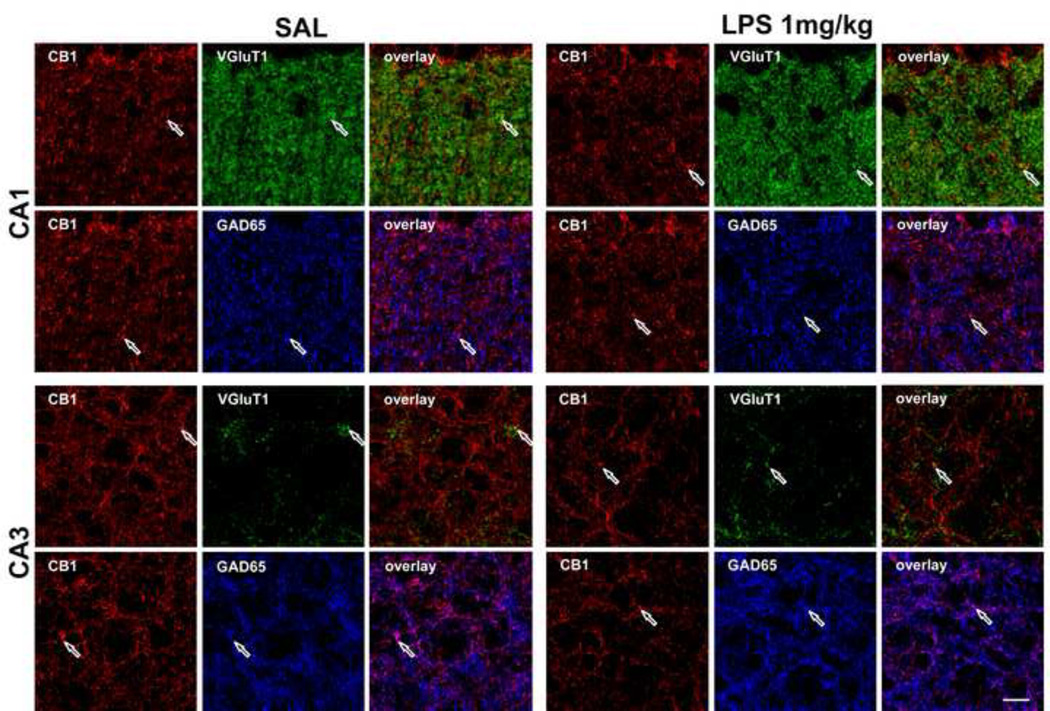
Figure 7.
Confocal images of the CB1 (red), VGluT1 (green) and GAD65 (blue) immunoreactivity in CA1 radiatum layer and CA3 pyramidal layer of the hippocampus. CB1 immunoreactivity was relatively low in both regions, especially in CA3 pyramidal layer, in the LPS-treated mice. The colocalization of CB1/VGluT1, and CB1/GAD65 was represented as yellow and purple colors, respectively in the overlay panel. Examples of specific sites of overlap are shown with arrows. Scale bar: 10 µm.

