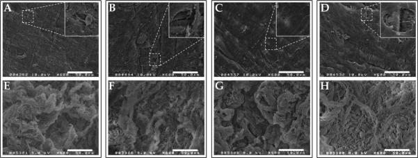
FIGURE 2.
Surface characterization of decellularized human umbilical vein scaffolds. Human umbilical veins (HUV) were decellularized using one of four chemical treatments: ethanol/acetone (A,E), sodium chloride (B,F), sodium dodecyl sulfate (C,G), or Triton X-100 (D,H). Shown are representative scanning electron micrographs of the lumenal (A–D) and ablumenal (E–H) surfaces. All four treatments resulted in complete removal of resident cells from the surfaces of the HUV, while leaving behind the basement membrane (A–D). Small areas where the underlying fibrillar collagen are exposed underneath denuded basement membrane are shown in the insets. Scale bar: 50 μm.

