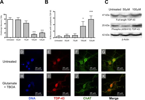
Figure 5. Glutamatergic toxicity induces the cytoplasmic translocation of TDP-43 and the selective death of motor neurons in vitro.
(A) Glutamate elicited a dose-dependent decrease in the number of ChAT positive neurons relative to GFAP positive glia. treatment groups when compared to untreated cells, F=20.21, df=4 and P<0.0001. (B) The number of neurons with cytoplasmic TDP-43 staining increased in a dose-dependent manner as the concentration of glutamate was increased, F=7.747, df=4 and P<0.0001. (C) Glutamate also elicited an increase in expression of full length TDP-43 and the 25kDa C-terminal Phospho(409/410) TDP-43 fragment in glutamate treated motor neuron culture samples. (D–K) Examples of untreated and glutamate treated cells (10 m TBOA and 100 m glutamate for 24 hours). (D,E) In untreated neurons the localization of TDP-43 was restricted to the nucleus. (F,G) ChAT staining and the merged image of ChAT and TDP-43 are shown in untreated cells. (H,I) Following glutamate treatment, a portion of neurons had TDP-43 staining extending out of the nucleus and into the cytoplasm and dendritic arbors. (H–K) glutamatergic toxicity induced the cytoplasmic mislocalization of TDP-43 in ChAT positive neurons. *p< 0.05, compared to untreated using a Bonferonni post hoc analysis.

