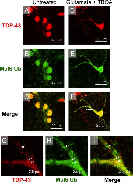
Figure 6. TDP-43 co-localizes with multi-ubiquitin following glutamate toxicity induced nucleo-cytoplasmic translocation.
(A–C) In untreated neurons TDP-43 immunostaining was restricted to the nucleus. Staining for multi-ubiquitin was also mainly nuclear with some pucnta appearing in the cytoplasm. (D–F) Following glutamate treatment, TDP-43 and multi-ubiquitin were both translocated from the nucleus into the cytoplasm. (G–I) High magnification images of inset from panel F show glutamate-induced cytoplasmic co-localization of TDP-43 and multi-ubiquitin (white arrows).

