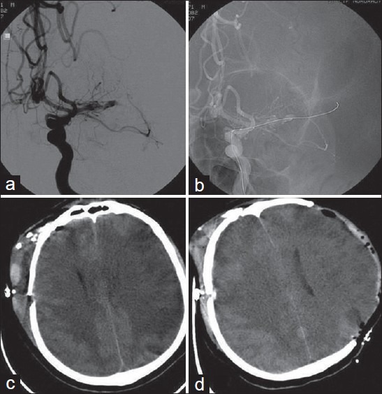
Figure 1.

A case of SAH due to ruptured right MCA bifurcation aneurysm. Malignant vasospasm was developed in the territory of the left MCA (a) Cerebral angiography after surgical clipping of the aneurysm and developement of the vasospasm showing severe vasospasm in the left MCA (b) Cerebral angiographic views during the attempt of angioplasty of the left MCA (c) Brain CT-scan showing ischemic changes and edema in the contralateral MCA territory with midline shift. (Note: right intraventricular catheter is not visible as this CT section is immediately below the tip of the catheter which slightly upward migrated after subfalcian herniation) (d) Brain CT-scan obtained after decompressive craniectomy performed via a left fronto-parietotemporal craniectomy and removal of the intraventricular catheter
