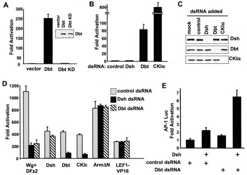
FIG. 7.
Function of Dbt and Dsh during β-catenin and JNK/planar cell polarity signaling in Drosophila S2 cells. (A) Overexpression of Dbt activates Wnt signaling in S2 cells; 0.2 μg of empty vector or Myc-tagged Dbt cDNA in a D. melanogaster expression plasmid was cotransfected with 0.1 μg each of Drosophila expression construct with the coding sequence for LEF-1, Renilla luciferase, and LEF-1 luciferase reporter. The experiments were done in triplicate, and the luciferase activities were normalized to Renilla luciferase activity. The Dbt KD mutant contains a K38D mutation in the kinase domain and lacks kinase activity. Expression of Myc-tagged Dbt and Dbt KD mutant in S2 cells was confirmed by immunoblotting with anti-Myc antibodies (inset). (B) The effects of Dbt-dsRNA and CKIα-dsRNA on basal LEF-1 transcription. S2 cells were plated into 12-well plates and treated with the indicated dsRNAs at a concentration of 15 μg/well. Twenty-four hours after dsRNA treatment, cells were transfected with the LEF-1 reporter plasmids. Cells were lysed 36 h after transfection, and luciferase activities were measured. (C) Effectiveness and specificity of Dsh-, Dbt-, and CKIα-dsRNA. S2 cells were treated with the indicated dsRNA and transfected with Myc-tagged Dsh, Dbt, or CKIα expression constructs. Thirty-six hours after transfection, cells were lysed and subjected to Western blotting with anti-Myc antibodies. (D) The effects of Dbt-dsRNA and Dsh-dsRNA on activated LEF-1-mediated transcription. S2 cells were plated into 12-well plates and treated with the indicated dsRNAs at a concentration of 15 μg/well. Twenty-four hours after dsRNA treatment, cells were transfected with the indicated effector plasmid together with LEF-1 reporter plasmids. Cells were lysed 36 h after transfection, and luciferase activities were measured. (E) The effects of Dbt-dsRNA on Dsh-dependent JNK signaling. S2 cells were treated with control or Dbt-dsRNA and then transfected with a Dsh expression plasmid together with an AP-1 luciferase reporter plasmid and a cytomegalovirus-Renilla luciferase reporter plasmid. Cells were lysed 48 h after transfection, and luciferase activities were measured.

