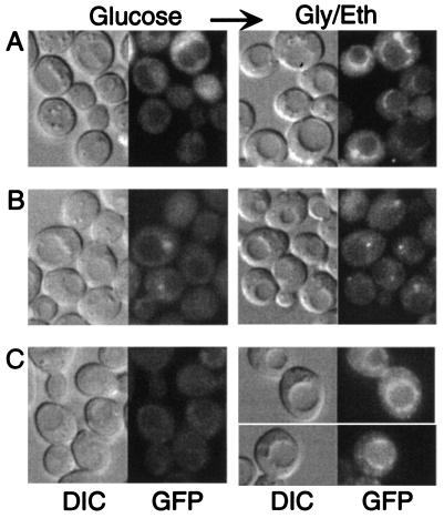
FIG. 2.
Snf1 accompanies Sip1 to the vacuolar periphery but is not required for localization. Transformants of strains MCY4097 (sip2Δ gal83Δ) (A) and MCY4098 (sip1Δ sip2Δ gal83Δ) (B) expressed Snf1-GFP from pOV84. (C) MCY4908 (snf1Δ) cells expressed Sip1-GFP from pOV90. Cells were grown in glucose and shifted to glycerol-ethanol (Gly/Eth) for 30 min. The arrow indicates a shift. GFP fluorescence and differential interference contrast (DIC) images are shown.

