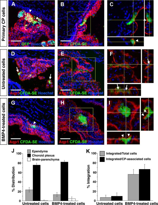
Figure 5.

Engraftment of endogenous CP by primary and derived CPECs following intraventricular injection. A–C, Primary CP cell injections, single or collapsed z-stack images of ipsilateral telencephalic choroid plexus 2 days after injection. Injected H2B-GFP (A) or CFDA-SE-labeled CP cells (B) preferentially associate with host CP, which expresses Aqp1 (red). Confocal projections reveal labeled cells on the nuclear side of the continuous Aqp1-positive apical border (C, arrowheads), indicating integration of injected cells into host CP with appropriate apicobasal polarity. D–F, Mouse ESC-derived (M1) aggregates, no BMP4 treatment, 2 d after injection. Dye-labeled cells are associated with ipsilateral host CP, but remain on the ventricular side of the Aqp1-expressing apical border (F, arrows), indicating a failure to engraft host CP. G–I, Mouse ESC-derived (M1) aggregates, with BMP4 treatment and Ara-C enrichment, 1–2 d after injection. Like the primary cells, most of the dye-labeled cells associate with host CP and are appropriately polarized on the nuclear side of the endogenous Aqp1 border (G, I, arrowheads), indicating host CP engraftment. J, K, Quantification of distributions and integration of injected BMP4-treatedand-untreatedcells. Cells from both groups are found mostly in association with CP (J), but the BMP4-treated cells integrate into host CP with much greater efficiency (K), consistent with their dCPEC identity (see Results for numbers; error bars represent SEM.). Scale bars, 50 μm.
