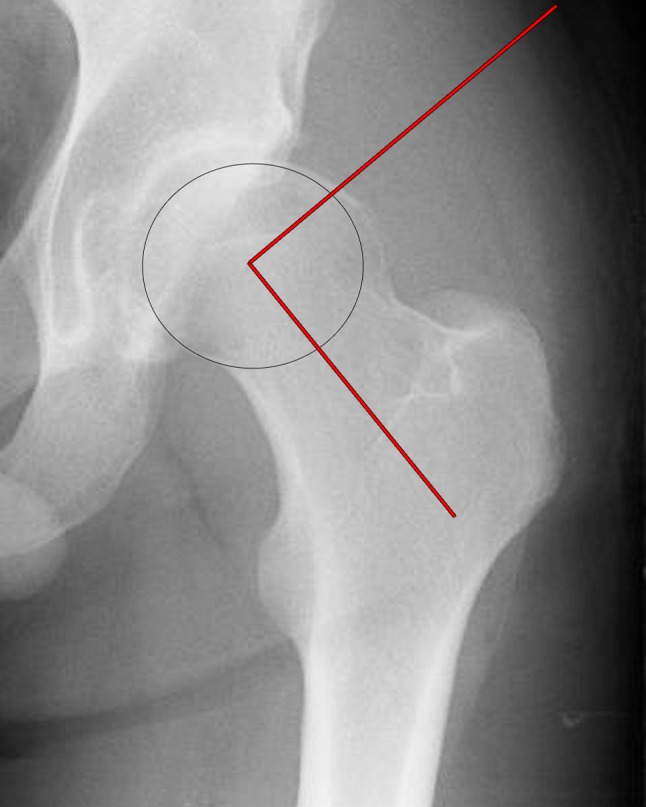
Fig. 1.

A magnified anterior posterior radiograph view taken from a pelvis radiograph with an increased alpha angle. The alpha angle subtended between a line from the midline of the femur to the center of the femoral head and a line from the center of the femoral head to the point at which the femoral head deviated from a circular template overlay
