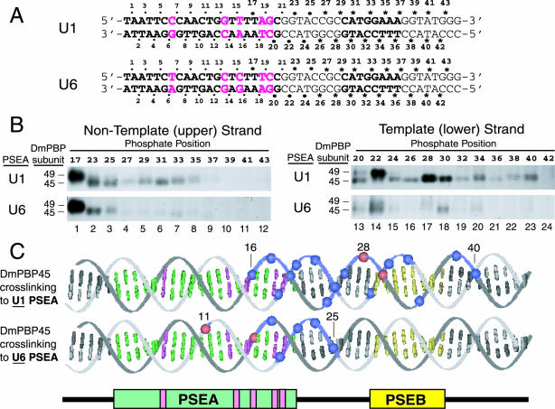
FIG. 1.
Site-specific protein-DNA photo-cross-linking of DmPBP to phosphate positions downstream of the PSEAs of D. melanogaster U1 and U6 genes. (A) Sequences of the relevant areas of the U1 and U6 photo-cross-linking probes. The sequences of the 21-bp U1 and U6 PSEAs are shown in boldface type. (Also shown in boldface type is the sequence of an 8-bp U1 PSEB, which is a second sequence fairly well conserved in D. melanogaster snRNA genes transcribed by RNA polymerase II [16, 38].) The five base pairs where the U1 and U6 PSEAs differ are indicated by magenta letters. Except for these five positions, the probes were identical in sequence to ensure that any differences in the cross-linking pattern were due solely to the five differences within the two PSEAs. The individual phosphate positions at which a cross-linker was incorporated are indicated by the dots, asterisks, and numbers above and below the sequences. The dots indicate positions for which data were reported in an earlier study (33). The asterisks indicate new positions not previously investigated; these 36 phosphate positions were individually derivatized to generate the 36 new probes used in this study. The larger dots indicate positions repeated in the present study as intensity standards and size markers to relate the new studies to the previous one. (B) Twenty-four different radiolabeled site-specific U1 probes and 24 different U6 probes were each incubated in separate reaction mixtures with DmPBP. Following UV irradiation and nuclease digestion, polypeptides that cross-linked to the DNA were detected by SDS-gel electrophoresis and autoradiography. Only the regions of the gels that correspond to the mobilities of DmPBP49 and DmPBP45 are shown. The left panels show the results of cross-linking to the nontemplate strand, and the right panels show cross-linking to the template strand. In each case, the upper panel illustrates the pattern for the U1 PSEA and the lower panel illustrates the pattern for the U6 PSEA. (C) Summary of DmPBP45 cross-linking to DNA probes that contain either the U1 PSEA or the U6 PSEA. Cross-linking data from Fig. 1B, together with previously reported data (33), are shown projected onto B-form DNA. Phosphate positions that cross-linked relatively strongly to the 45-kDa subunit of DmPBP are indicated by blue spheres, whereas the very strongest cross-linking positions are indicated by red spheres. Weak cross-links are not indicated. Base pairs that comprise the PSEA are shown in green and magenta, and those that comprise the U1 PSEB are shown in yellow. When DmPBP was bound to a U1 PSEA, phosphate positions between 16 and 40 cross-linked to DmPBP45, but when DmPBP was bound to a U6 PSEA, cross-linking to DmPBP45 was restricted to a region between phosphates 11 and 25. When the DNA is oriented as shown, the cross-links to DmPBP45 are mainly on the upper surface of the DNA helix.

