Fig 3.

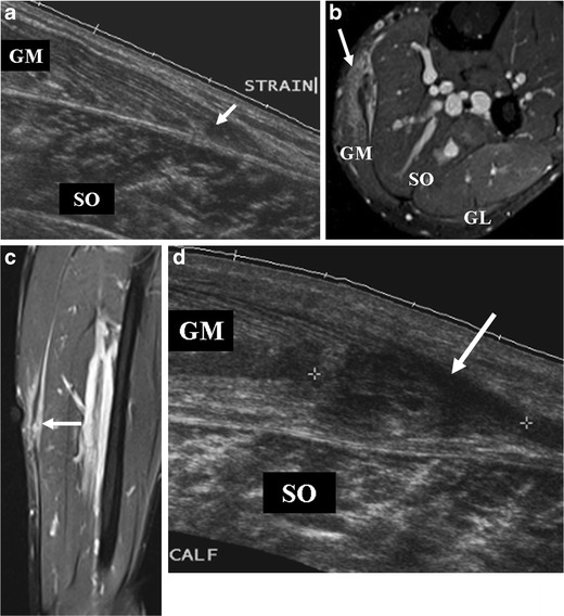
A 27-year-old football player presenting with left calf pain, clinically diagnosed as grade 2 injury. a On ultrasound at initial presentation, a hypoechoic area measuring 1.0 × 0.4 cm was noted (arrow), corresponding with a partial tear of the medial head of the gastrocnemius (GM). Colour Doppler imaging was normal (not shown). No evidence of haematoma or other abnormality was observed. Soleus muscle (SO) appears intact. MR imaging was not performed on this patient at this time. b-d Follow-up imaging 2 months after initial presentation. b Axial and (c) coronal FS T2-w TSE images reveal areas of hyperintensity with feathery appearance, consistent with a partial tear of the medial head of the gastrocnemius muscle with intramuscular oedema (arrow). Note the peritendinous oedema. d On ultrasound, a persistent hypoechogenic area was noted that appeared to have increased in extent (arrow). Colour Doppler imaging was normal (not shown). A 7-month follow-up MR imaging showed complete recovery (not shown)
