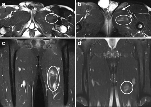
Fig 6.

A 19-year-old gymnast presenting with an injury of the left groin. Axial FS T2-w TSE image shows (a) abnormal hyperintensity (oval) consistent with a strain of pectineus muscle, (b) discrete hyperintensity within the adductor longus (arrow, strain without a tear) and mild haematoma within the adductor brevis (oval, partial tear). (c) Haemorrhagic fluid extending into the fascia surrounding the adductor longus is shown (oval). The area of perifascial fluid spans a length of 10.8 cm over the proximal to mid aspects of the thigh. (d) The MR imaging at the 1-month follow-up showed some residual hyperintensity in the medial thigh (circle) corresponding with persistent oedema surrounding the adductor longus, but near-complete resolution of haematoma is noted. Normal homogeneous signal intensity was seen within the pectineus, adductor longus and brevis muscles, indicating recovery (not shown)
