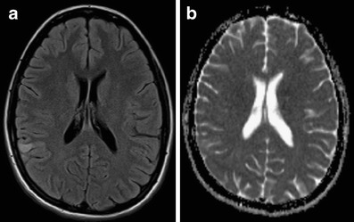
Fig. 2.

A 20-year-old female with sudden onset of neurological symptomatology. a FLAIR image shows an area of increased signal intensity in the right angular region. b ADC map does not show restricted diffusion that would be characteristic for an acute ischemic stroke. Encephalitis was proven by CSF examination. FLAIR, fluid attenuated inversion recovery; ADC, apparent diffusion coefficient; CSF, cerebrospinal fluid
