Fig. 7.

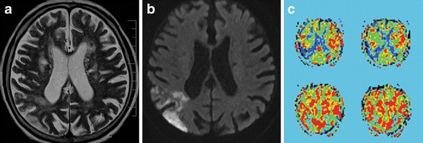
An 86-year-old male with a history of previous ischemic events developed sudden left-sided hemianopia. a T2-weighted TSE image with a number of ischemic lesions. b DW image (b = 1,000) clearly shows a region of acute ischemia as an area of restricted diffusion. c PASL image with a perfusion deficit in the right occipital region. TSE, turbo spin echo; DW, diffusion weighted; PASL, pulsed arterial spin labeling
