FIG. 4.

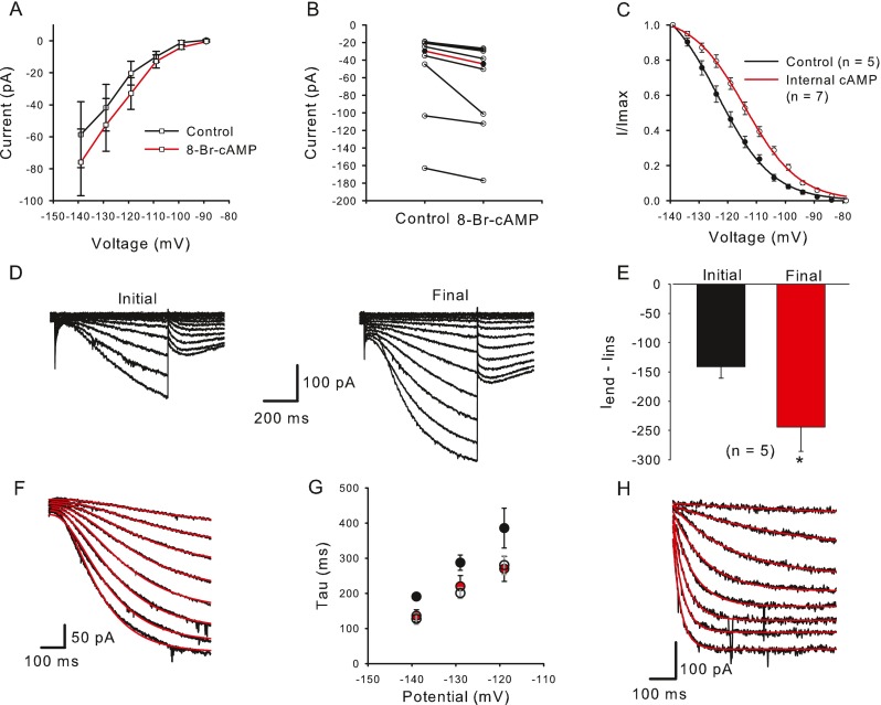
Cyclic AMP and GMP increase I h and shift its activation. A In the presence of external 8-Br-cAMP (data points connected by red line) mean I end − I ins was larger at each voltage step compared to controls (black line). The mean current amplitude increased from −58.6 to −76.0 pA during a step to −139 mV (n = 7). B Current values for eight cells are shown for control and 8-Br-cAMP. Median current amplitude (red line, I end − I ins) increased significantly from −29.9 to −44.4 pA at the −139 mV voltage step (P = 0.008, n = 8, Wilcoxon signed rank test). C Activation curves were obtained from control cells (no internal cAMP, black line) and in the presence of internal cAMP (red line). I/I max was obtained from normalized tail currents and the means ± SEM were plotted against prepulse voltage for controls (closed circles) and cAMP (open circles) and fitted with a Boltzmann function (Eq. 2). V 1/2 obtained from a fit to the mean data shifted from −123.4 mV (n = 5) to −113.9 mV (n = 7) with internal cAMP. D Whole-cell currents increased in the presence of 200 μM internal cGMP. E Mean current amplitude increased from an initial value of −141.3 to −244.1 pA after dialysis with cGMP. Control currents were measured within 2.5 min after breakthrough to the cell and final currents represent the maximum current reached. F, G cAMP and cGMP speed activation kinetics of I h. F Representative current traces (black) to voltage steps from −144 to −109 mV (5 mV increments). The activation phase of I h was fit with a single exponential with third order kinetics (Eq. 3) (red lines). G Mean time constants (tau) of activation for control cells (black circles, 14–28 cells) and cells exposed to internal cAMP (red circles, 3–5 cells) or cGMP (unfilled circles, 4 cells) for three different voltage steps. The time constant at the −139 mV step was significantly faster in the presence of internal cAMP (P = 0.029; n control = 28; n cAMP = 5) and internal cGMP (P = 0.013; n cGMP = 4), Mann–Whitney rank sum test. H Activation kinetics are faster in a type II hair cell. Voltage steps were from −164 to −94 mV (10 mV increments). Current activation was fit with a single exponential with first order kinetics (red lines). Tau for a step to −144 mV was 51.1 ms.
