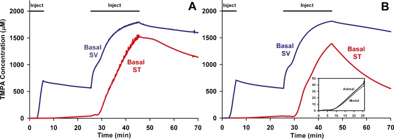
FIG. 1.
In vivo recordings of TMPA concentration during lateral semi-circular canal (LSCC) injections. A: TMPA concentration measured simultaneously from ion-selective electrodes sealed into ST and SV of the basal turn during two injections (of 7 min and 20 min duration) of 2 mM TMPA solution into the lateral canal at 1 μL/min. B: Computer simulation of the experiment in an anatomically-based model, with injections driving volume flow from the LSCC injection site towards the cochlear aqueduct through the perilymphatic spaces of the lateral canal, vestibule, scala vestibuli and scala tympani. The inset figure shows the initial measured and calculated time courses in ST shown enlarged. The increase of ST concentration during the initial period when SV concentration was elevated was used to quantify the rate of local cross-communication between ST and SV in the basal turn.

