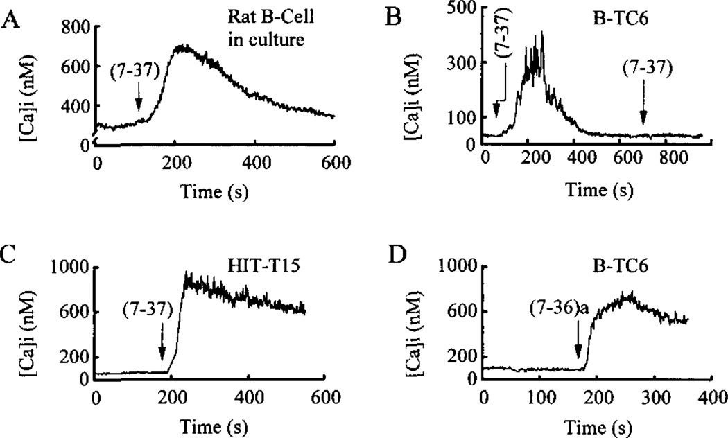
Fig. 1. GLP-1 increases [Ca2+]i, in rat β-cells and pancreatic insulinoma cells.
Each of the two isoforms of GLP-1, abbreviated as (7-37) or (7-36)a, were administered to individual cells at a concentration of 10 nm for 10 s (arrows indicate the start of application). The GLP-1 was then removed within 30 s by a superfusion system. A sustained rise of [Ca2+]i in response to GLP-1 was observed in a rat β-cell maintained in primary cell culture (panel A), in βTC6 cells (panels B and D), and an HIT-T15 cell (panel C). In panel A the extracellular solution contained 7.5 mm glucose, whereas in panels B-D it contained 0.8 mm glucose.

