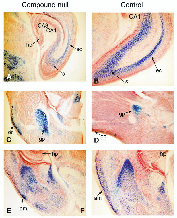
FIG. 4.
Histological comparison of brain morphology in compound null Lmo1 or Lmo3 mutant mice. Sections of various brain regions from (Lmo1−/−; Lmo3Z/Z) (designated compound null) or (Lmo1+/−; Lmo3Z/+) (designated control) mice are compared. Brains were removed from mice transcardially perfused with 4% PFA at P0, and sections were made on a sliding microtome. These were stained free-floating with X-Gal, mounted, and counterstained with neutral red. Regions of brain illustrated in sections shown are abbreviated as follows: hp, hippocampus; s, subiculum; ec, entorhinal cortex; oc, olfactory cortex; gp, globus pallidus; am, amygdala; CA1, CA3 of hippocampus.

