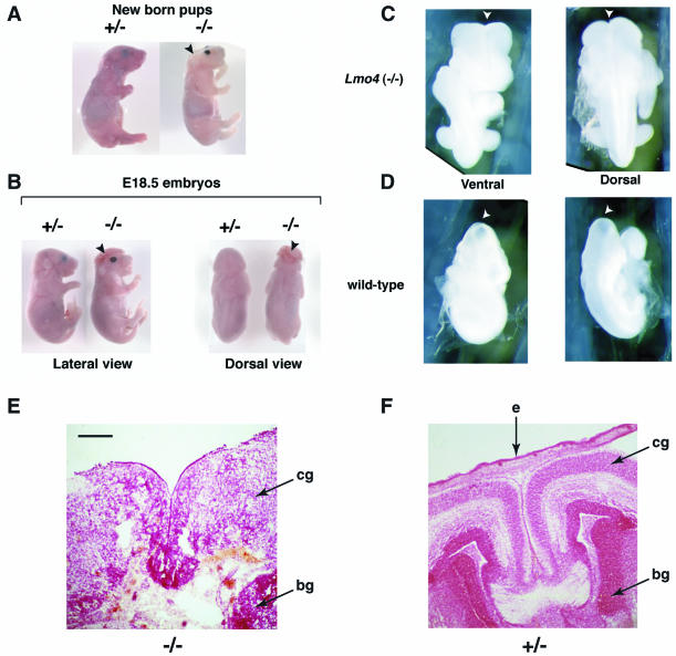
FIG. 6.
Neural tube closure defect in Lmo4 homozygous null mutant mice. Timed matings between heterozygous Lmo4 mice were terminated at P0 (A), E18.5 (B), or E9.5 (C and D), and pups were genotyped by filter hybridization using the 3′ probe. (A to D) Whole-mount photographs are shown. (A) Lateral view of a homozygous Lmo4−/− dead-born pup, compared to a heterozygous Lmo4+/− live-born littermate. The Lmo4−/− pup exhibits anencephaly in which the cranium and the brain are absent. The facial features are also markedly abnormal compared to those of the Lmo4+/− pup. (B) Lateral and dorsal views comparing an E18.5 homozygous Lmo4−/− embryo with a heterozygous Lmo4+/− littermate. The Lmo4−/− embryo exhibits exencephaly in which the cranium is absent and the is brain exposed. The nasal part of the Lmo4−/− embryo is broader, and the ears are located at a much lower position. (C and D) Posterior views of an E9.5 Lmo4−/− embryo show that the cranial neural tube remains open at the position of the anterior neuropore (arrowhead). The same views taken from a wild-type littermate reveal that the cranial neural tube has closed by E9.5. (E and F) Histology of dorsal telecephalon of homozygous or heterozygous Lmo4 embryos at E18.5. Coronal section through the head, showing complete loss of brain structure, necrosis, and loss of tissue (skin and mesenchyme) overlying the dorsal part of the brain in an Lmo4 homozygous embryo (E) compared with a heterozygous littermate (F). The brain stem in Lmo4 homozygous embryos was intact and apparently normal. Abbreviations: e, epidermis; cg, cortical grey matter; bg, basal ganglia. The sections were stained with neutral red. Scale bar: 100 μm.

