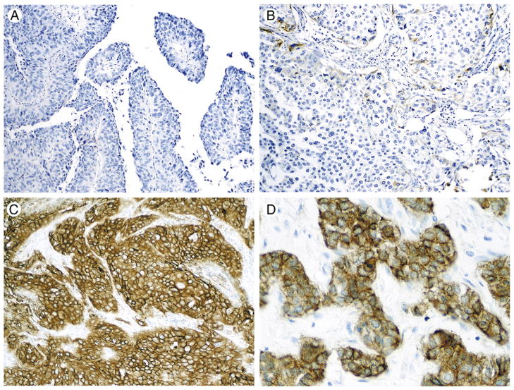
Fig. 1.
Patterns of EGFR immunoexpression. A, Invasive high-grade papillary urothelial carcinoma with no EGFR expression. B, Invasive high-grade urothelial carcinoma with focal positivity; cases that showed this staining pattern were considered as “EGFR positive, low expression level.” C, High-grade invasive urothelial carcinoma with strong and diffuse EGFR immunoexpression. Cases showing this staining pattern were considered as “EGFR positive, high expression level.” D, A higher magnification is depicted here.

