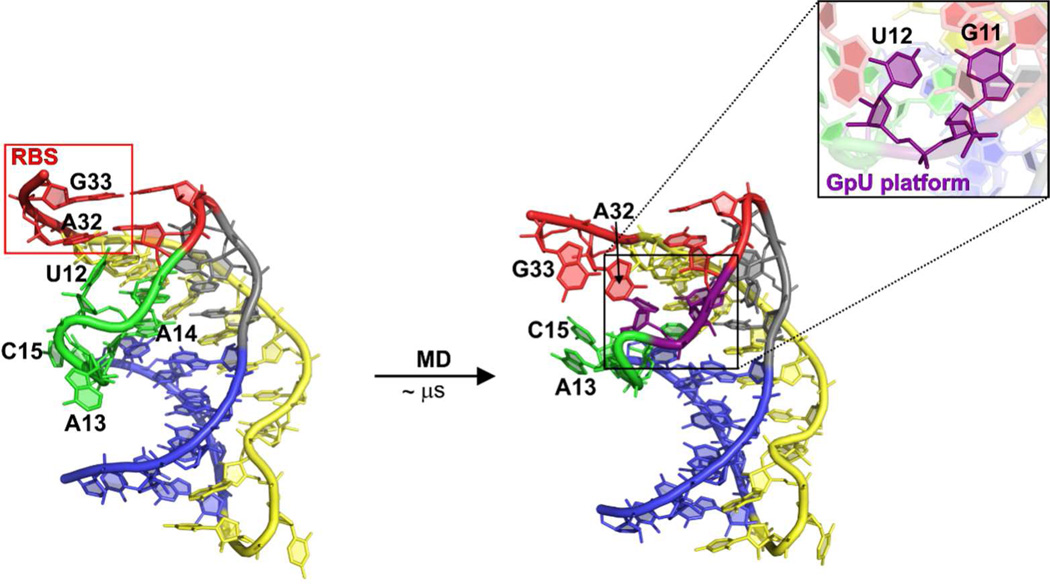
Figure 6.
Starting X-ray (left) and functional “ON” state (right) geometry of the ligand-free PQA taken from Q51-free-2 simulation. The GpU platform, which is formed by bases U12 and G11, is highlighted in the context of the whole “ON” state PQA as well as in the close-up using the violet sticks. Note that the ribosome binding site (RBS), including bases A32 and G33, becomes considerably less structured in the “ON” state ligand-free PQA structure.

