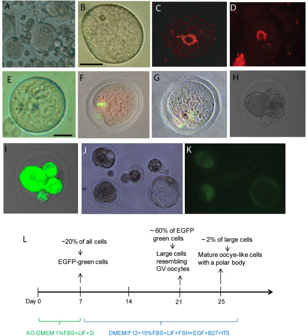
Figure 2 .
Characterization of mature oocytes and embryos derived from spermatogonial stem cells (SSCs) in culture.(A and B) Phase contrast microscopy of growing oocytes from the culture of SSCs (A), a SSCs-derived oocyte (SSC-Ooc) resembling that of germinal vesicle (GV) stage (B,scale bar = 30 μm). (C and D) Hoechst staining of a GV oocyte from a mouse ovary (C) and OG2-SSC-Ooc (D) showing a rim of chromatin around the nucleolus (the surrounded nucleolus). (E) Phase contrast microscopy of a fully-grown SSC-Ooc, scale bar = 30 μm. (F and G) YoYo1(green) and lamin B1 (red) staining of a control MII oocyte from a normal mouse ovary (F) and an oocyte with a polar body derived from SSC (G). (H and I) A 4-cell embryo was generated from SSC-oocyte by ICSI with sperm from an OG2 mouse, from which GFP gene was carried by sperm and was expressed in the resulting embryo (I). (J and K) Phase contrast microscopy of parthenogenetic embryos developed from OG2-SSC-oocytes in culture, Oct4/GFP from OG2 strain was expressed in the embryos (K). (L) Schematic representation of the reprogramming conditions from SSCs to oocyte-like cells.

