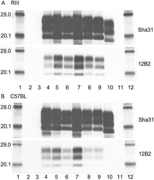
Figure 5.

Western blot analysis in wild‐type mice. Western blot analysis of proteinase‐K‐digested PrPSc from UK‐1 and UK‐2 challenged RIII (A) and C57BL (B) mice, detected with Sha31 and 12B2 antibodies. Lanes 1 and 12: molecular mass markers (kDa), Lanes 2 and 11: blank, lane 3: unchallenged proteinase‐K digested mouse brain homogenate, lane 4: mouse challenged with ovine classical scrapie, lanes 5–6 and 8–9: mice challenged with UK‐1 or UK‐2, lane 7: mouse challenged with murine passaged ME7 strain, lane 10: mouse challenged with sheep bovine spongiform encephalopathy (BSE). Exposure time was1 minute for all blots.
