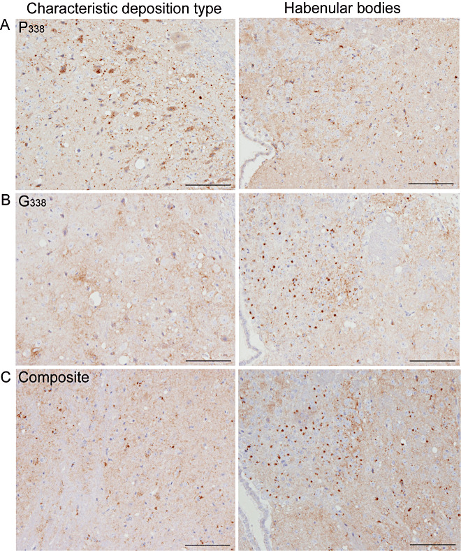
Figure 8.

PrPSc deposition patterns of UK‐1 following transmission to tg338 mice. PrPSc labeling in tg338 mice showed distinct patterns and deposition types. The characteristic deposition type and labeling within the habenular bodies are also shown. P338 (A) was predominantly punctuate and intraneuronal labeling. G338 (B) was characterized by granular labeling within the neuropil and distinctive aggregates within the medial habenular bodies. A number of mice showed a mixture of P338 and G338 (composite) (C). Scales bars represent 100 µm.
