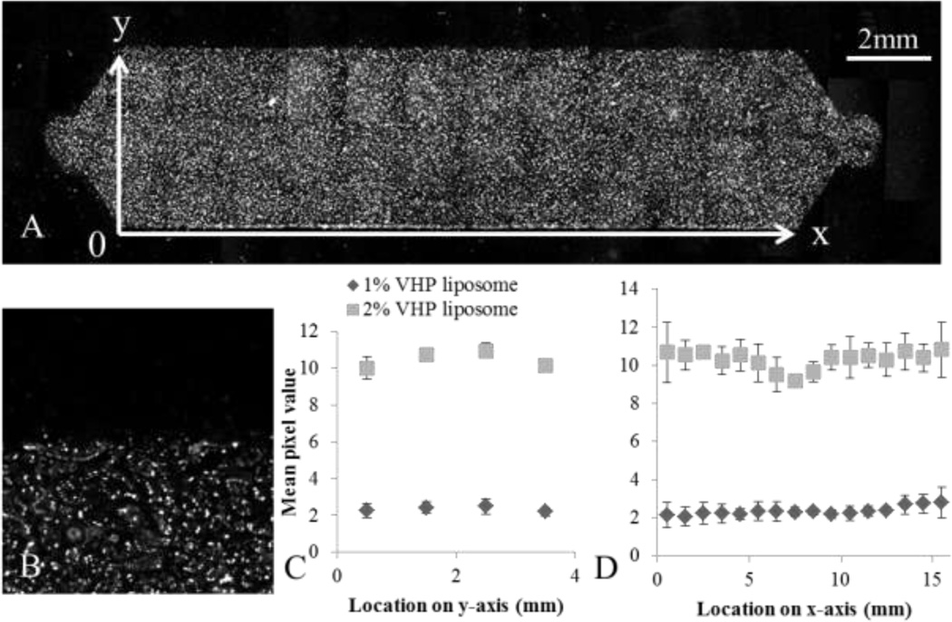
Figure 5.
Spatial binding pattern of liposomal nanoparticles throughout the USC was characterized by flowing 1 (N = 8) and 2 % (N = 6) VHP-conjugated liposomes over HUVEC and measuring fluorescence through microscopy. A) 20×6 frame mosaic image composed of fluorescence images acquired using a 5× objective, with background subtraction. B) An enlarged image of the edge of the chamber, showing the individual cells with punctate fluorescence, seen at 5×. C) The average fluorescence along the y-axis. D) The average fluorescence along the x-axis. One way ANOVA results showed significant spatial dependence of 2% VHP-conjugated liposomes capture along the y-axis (p < 0.01), but not along the x-axis. Capture of 1% VHP-conjugated liposomes was not spatially dependent along either axis (p > 0.01).

