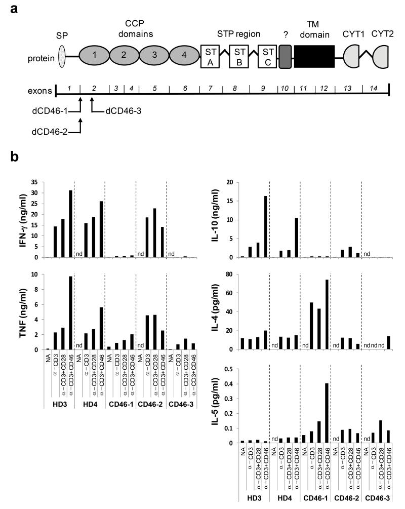
Figure. 5. T cells from CD46-deficient patients present with defective in vitro TH1 induction.
(a) Localization of the CD46 gene mutations in the three CD46-deficient patients (CD46-1 to CD46-3) assessed for TH1 induction. The lower part shows the exon structure of the CD46 gene and above the corresponding protein domains. (b) Comparison of cytokine expression by CD4+ T cells from healthy donors and CD46-deficient patients. T cells from two healthy donors (HD3 and HD4) or patients were purified from freshly-drawn blood samples and either left non-activated (NA), or activated with immobilized antibodies to CD3 and CD46 in the presence of 25 U/ml rhIL-2. Indicated cytokine secretion into the cell culture media was assessed 36h post activation using the TH1 and TH2 CBA Cytokine Secretion Assay. Data shown are the mean value of each condition performed in duplicate. Note, that though values for only two HD are shown, these are representative for 12 age- and gender-matched donors assessed over the course of the study. CCP, complement control protein; CYT1 or CYT2, cytoplasmic tail 1 or −2; nd, not detectable; SP, signal peptide; STP, serine threonine proline-rich; TM, transmembrane;?, region of unknown function

