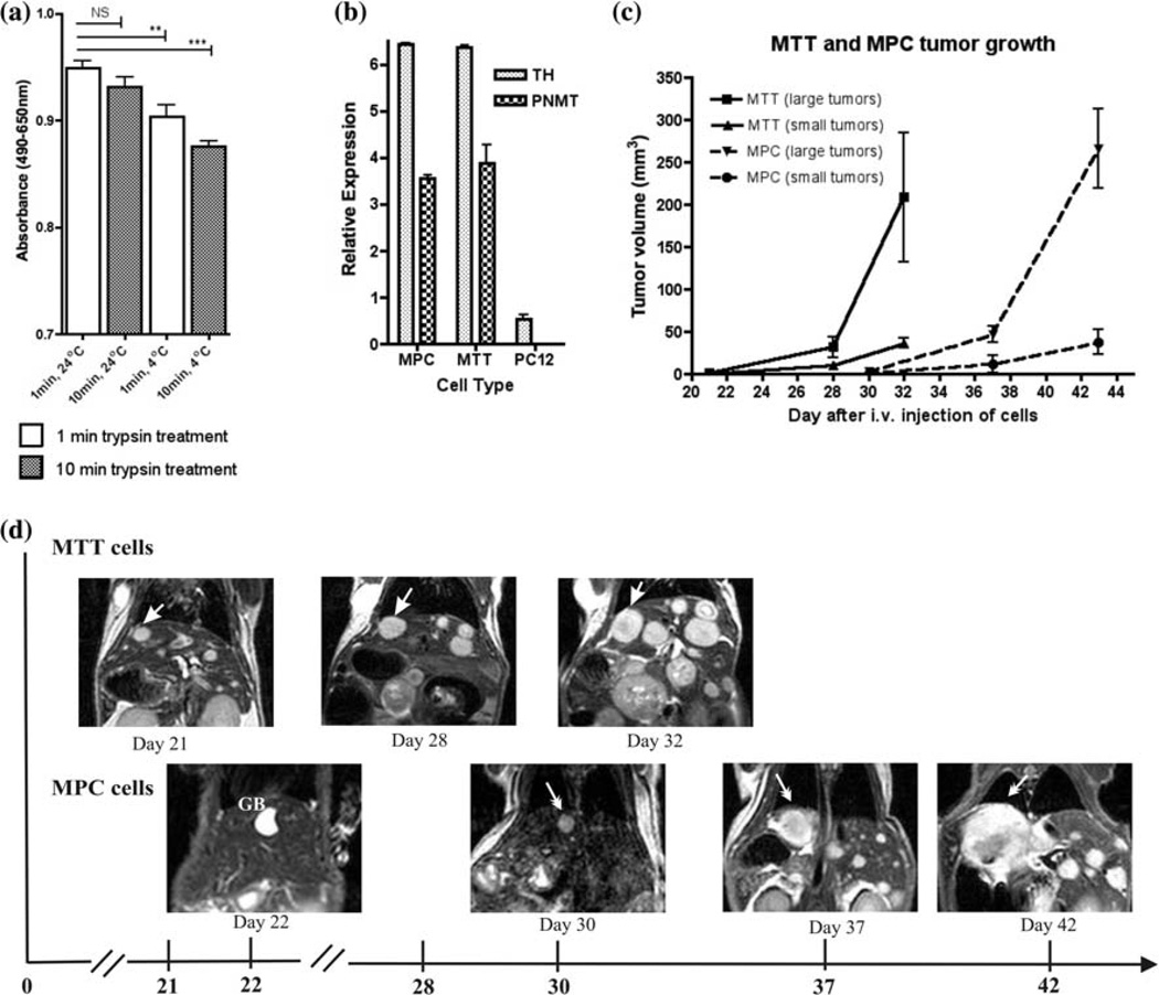
Fig. 1.
In vitro and in vivo characterization of MPC and MTT cells. a Cell proliferation assay showing the effect of different condition for MPC cells. Trypsin did not show a significant effect if the cells were treated at room temperature, but was significant at 4°C. An un-paired t-test analysis of these data with P value less than 0.01 as significant (*P < 0.01; **P < 0.001; ***P < 0.0001) was performed. b Expression of TH and PNMT mRNA in MPC, MTT, and PC12 cells determined by qRT-PCR relative to levels of 18S RNA. In vivo characterization of metastatic pheochromocytoma with MRI; c Small and large tumor growth in mice injected with MPC (n = 5) or MTT (n = 5). Mice injected with MTT developed tumors approximately 10 days earlier than mice injected with MPC. d Serial MRI images in same mouse scanned on days 21, 28, and 32 for MTT and on days 22, 30, 37 and 42 for MPC cells. Arrows indicate the same liver tumor

