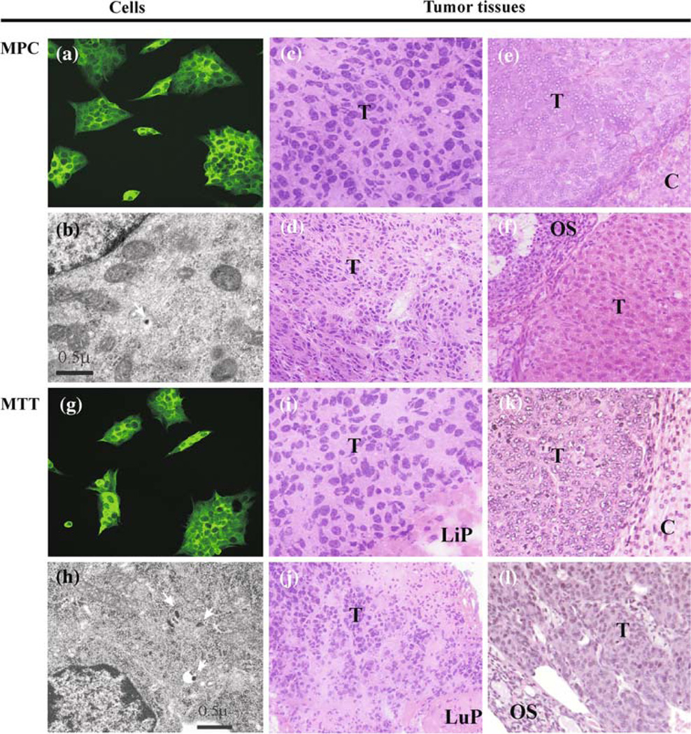
Fig. 2.
Characterization of MPC and MTT tumor histopathology. Immunocytochemistry reveals expression of TH in MPC (a) and MTT (g). Ultrastructure by electron microscopy reveals secretory dense cores granules (arrows) typically seen in pheochromocytomas in both cell lines (b, h); scale bar = 0.5 µm. Multiple organ lesions from MPC and MTT models: liver (ci); adrenal gland (ek); lung (dj); ovary (fl) reveal similar histopathology (H&E) suggestive of neuroendocrine tumor origin, magnification 40×. Elevated tissue catecholamine levels were demonstrated in these lesions confirming pheochromocytoma. Abbreviations T tumor, Li liver parenchyma, A adrenal cortex, Lu lung parenchyma, O ovarian Stroma

