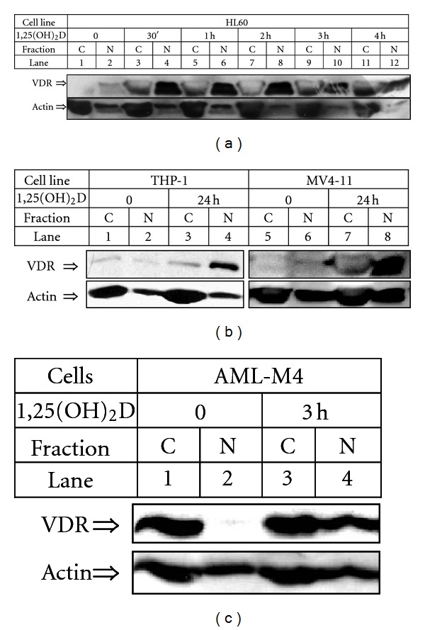
Figure 5.

Expression of VDR protein in AML cells exposed to 1,25(OH)2D. AML cells after incubation for indicated times with 10 nM 1,25(OH)2D were lysed and fractionated into the cytoplasmic (C) and nuclear (N) fractions. The lysates from equal numbers of cells were separated in SDS-PAGE and blotted to the membrane. The membrane was probed with anti-VDR. Actin was probed as a control of equal loading and transfer of proteins. (a) HL60 cells, (b) THP-1, MV4-11 cells, and (c) AML-M4 blasts from patient's peripheral blood. Based on [41, 42] and on unpublished data.
