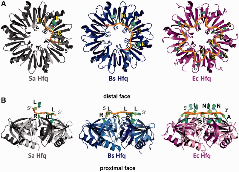
Figure 2.
The RNA-binding motifs of Hfq in Gram-positive and Gram-negative bacteria. (A) View looking down onto the distal face of Sa Hfq (left, colored grey), Bs Hfq (middle, colored blue) and Ec Hfq (right, colored magenta). Note the assigned colors will be used in all subsequent figures. Bound RNA is shown as a cartoon with sugar phosphate backbone colored yellow and the purine bases colored green. Each protein is labeled and colored appropriately. The purine nucleotide sites are labeled R, the linker sites are labeled L and the A-sites, which are found only in the Ec Hfq, are labeled A. The 5′- and 3′-ends of the RNA are labeled. (B) Side view of the Sa Hfq–A4 (left), Bs Hfq–(AG)3A (middle) and Ec Hfq–A15 (right) complexes. Each Hfq is labeled and colored as in (A) and the ‘distal’ and ‘proximal’ faces are labeled. Contiguous subunits are colored light and dark grey, blue or magenta in the respective complexes.

