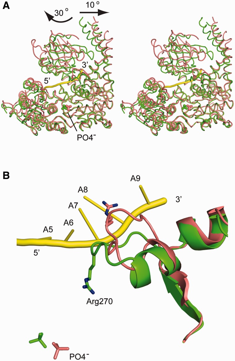
Figure 3.
Conformational changes of hIghmbp2hd upon RNA binding. (A) Stereo view showing the superposition of hIghmbp2hd–RNA (pink) with hIghmbp2hd (green). Phosphate ions are shown as sticks and the bound ssRNA is shown in yellow cartoon. The arrows indicate the movement of domain 1B and 1C in the RNA-bound state compared to the RNA-free state. (B) Conformational change of a loop region (residues 264–273) and the reorientation of residue Arg270 in response to ssRNA binding. The position of the phosphate ions indicates the location of the nucleotide-binding site.

