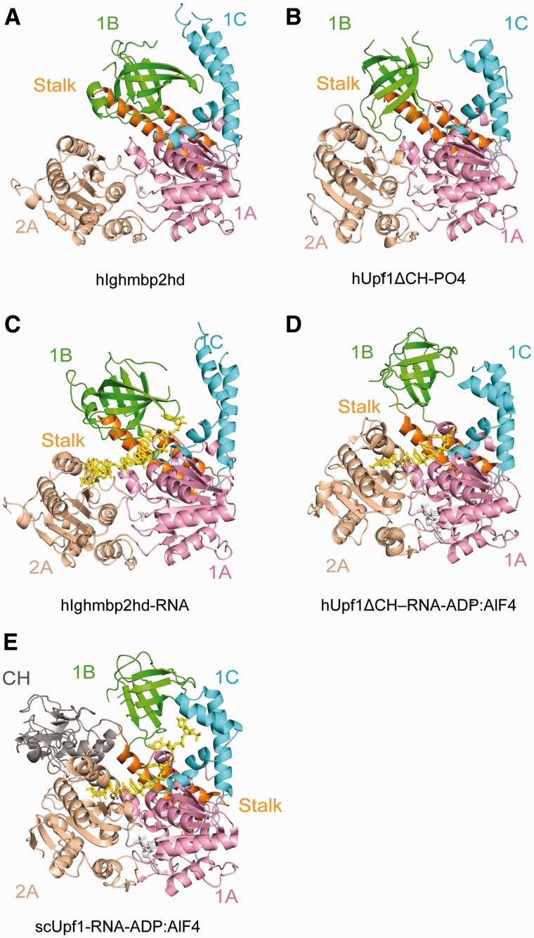
Figure 4.
Structural comparison of Ighmbp2 with Upf1. The ribbon diagrams are drawn with domains 1A (pink) and 2A (wheat) in the same orientation. The bound phosphate ion and nucleotide are shown in gray sticks. ssRNA is show in yellow sticks. The coloring scheme for domains are as in Figure 1. (A) hIghmbp2hd. (B) hUpf1ΔCH-PO4− (PDB code: 2GK7). (C) hIghmbp2hd–RNA. (D) hUpf1ΔCH-RNA-ADP:AlF4− (PDB code 2XZO). (E) Yeast Upf1-RNA-ADP:AlF4− (PDB code: 2XZL).

